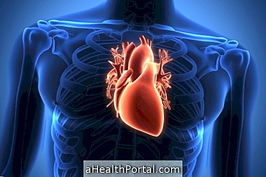
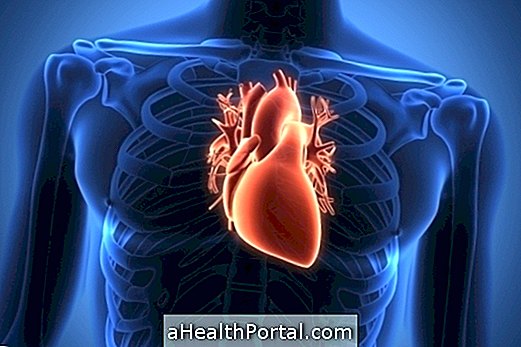
Mitral stenosis corresponds to the thickening and calcification of the mitral valve, resulting in the narrowing of the opening that allows the passage of blood from the atrium to the ventricle. The mitral valve, also known as the bicuspid valve, is a cardiac structure that separates the left atrium from the left ventricle.
According to the degree of thickening and, consequently, orifice size for blood passage, mitral stenosis can be classified into:
- Mild mitral stenosis, whose opening for the passage of blood from the atrium to the ventricle is between 1.5 and 4 cm;
- Moderate mitral stenosis, whose opening is between 1 and 1.5 cm;
- Severe mitral stenosis, whose opening is less than 1 cm.
Symptoms usually begin to appear when the stenosis is moderate or severe, as blood flow becomes difficult, resulting in shortness of breath, easy tiredness and chest pain, for example, it is necessary to go to the cardiologist for confirmation the diagnosis and started the treatment.

Symptoms of mitral stenosis
Mitral stenosis usually does not present symptoms, however there may be some development after physical exertion, for example:
- Easy fatigue;
- Sensation of shortness of breath, especially at night, having to sleep sitting or lying down;
- Dizziness when getting up;
- Chest pain;
- Blood pressure may be normal or decreased;
- Pink face.
In addition, the person may feel their own heartbeat and cough with blood if there is rupture of the vein or capillaries of the lung. Know the main causes of coughing up blood.
Main causes
The main cause of mitral stenosis is rheumatic fever, a disease mainly caused by the bacterium Streptococcus pneumoniae, which causes inflammation in the throat, causes the immune system to produce autoantibodies, which leads to inflammation of the joints and, possibly, changes in cardiac structure. Here's how to identify and treat rheumatic fever.
Less commonly, mitral stenosis is congenital, that is, it is already born with the baby, and can be identified in exams performed soon after birth. Other causes of mitral stenosis that are rarer than congenital stenosis are: systemic lupus erythematosus, rheumatoid arthritis, Fabry's disease, Whipple's disease, amyloidosis, and a tumor in the heart.

How is the diagnosis made?
The diagnosis is made by the cardiologist through the analysis of the symptoms described by the patient, as well as some tests, such as chest X-ray, electrocardiogram and echocardiogram. See how it works and how the echocardiogram is done.
In addition, in the case of congenital mitral stenosis, the doctor can make the diagnosis from the auscultation of the heart, in which a heart murmur characteristic of the disease can be heard. Here's how to identify the heart murmur.
How to treat
The treatment for mitral stenosis is done according to the recommendation of the cardiologist, and individualized doses of the medications are indicated according to the patient's need. Treatment is usually done with the use of beta-blockers, calcium antagonists, diuretics and anticoagulants, which allow the heart to function properly, relieve symptoms and prevent complications.
In more severe cases of mitral stenosis, it may be recommended by the cardiologist to perform surgery for repair or replacement of the mitral valve. Find out about the postoperative period and recovery from heart surgery.
Possible Complications
As in mitral stenosis there is difficulty in the passage of blood from the atrium to the ventricle, the left ventricle is spared and remains at its normal size. However, as there is a large accumulation of blood in the left atrium, this cavity tends to increase in size, which may facilitate the appearance of cardiac arrhythmias, such as atrial fibrillation. In these cases, the patient may require the use of oral anticoagulants to reduce the risk of stroke.
Also, since the left atrium receives blood from the lungs, if there is a blood pool in the left atrium, the lung is having difficulty sending blood to the heart. Thus, the lung eventually accumulates a lot of blood and consequently can become soaked, resulting in acute pulmonary edema. Learn more about acute pulmonary edema.


