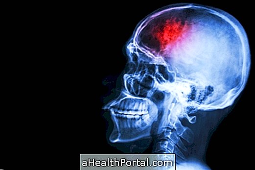
Angiotomography is a quick diagnostic exam that allows the perfect visualization of fat or calcium plaques inside the veins and arteries of the body, using modern 3D equipment, very useful in coronary and cerebral disease, but which can also be requested to evaluate vessels blood in other parts of the body.
The doctor who usually orders this test is the cardiologist to assess the impairment of the blood vessels in the heart, especially if there are other abnormal tests such as stress testing or scintigraphy, or for an assessment of chest pain, for example.

What is it for
Angiotomography serves to clearly observe the internal and external part, diameter and involvement of blood vessels, clearly showing the presence of calcium or fat plaques in the coronary arteries, and also serves to clearly visualize cerebral blood flow, or in any other area of the body, such as the lungs or kidneys, for example.
This test can detect even the smallest coronary calcifications resulting from the accumulation of fatty plaques inside the arteries, which may not have been identified in other imaging tests.
When can be indicated
The following table indicates some possible indications for each type of this exam:
- in case of symptoms of heart disease
- individuals with installed heart disease
- suspected coronary calcification
- to verify stent efficacy after angioplasty
- in case of Kawasaki disease
- evaluation of obstruction of the cerebral arteries
- cerebral aneurysm research evaluation of vascular malformations.
- evaluation of cerebral vein obstruction due to extrinsic causes, thrombosis
- evaluation of vascular malformations
- pre-ablation of atrial fibrillation
- post-ablation of atrial fibrillation
- evaluation of vascular diseases
- before or after placing a prosthesis
- vascular diseases
- pre and post evaluation of prostheses
- for the evaluation of vascular diseases
How the exam is done
To perform this exam, a contrast is injected into the vessel to be visualized, and then the person must enter a tomography machine, which uses radiation to generate images that are seen on the computer. Thus, the doctor can assess how the blood vessels are, whether they have calcified plaques or if the blood flow is compromised somewhere.
Necessary preparation
Angiotomography takes an average of 10 minutes, and 4 hours before it is performed, the individual should not eat or drink anything.
Medicines for daily use can be taken at the usual time with little water. It is recommended not to take anything that contains caffeine and no erectile dysfunction medication for up to 48 hours before the test.
Minutes before the angiotomography, some people may need to take a medication to decrease the heart rate and another to dilate the blood vessels, in order to improve their visualization of the cardiac images.
Was this information helpful?
Yes No
Your opinion is important! Write here how we can improve our text:
Any questions? Click here to be answered.
Email in which you want to receive a reply:
Check the confirmation email we sent you.
Your name:
Reason for visit:
--- Choose your reason --- DiseaseLive betterHelp another personGain knowledge
Are you a health professional?
NoMedicalPharmaceuticalsNurseNutritionistBiomedicalPhysiotherapistBeauticianOther


