Molar pregnancy, also called spring pregnancy or hydatidiform spring, is a rare condition that occurs during pregnancy due to changes in the uterus, caused by the multiplication of abnormal cells in the placenta.
This condition can be partial or complete, depending on the size of the abnormal tissue in the uterus and has no definite cause, but it can occur mainly because of the fertilization of two sperm in the same egg, causing the fetus to have only cells from the father .
The abnormal tissue that grows in the uterus looks like grape clusters and generates malformation in the placenta and fetus, causing a miscarriage and, in rare cases, the cells of this tissue spread and lead to the development of a type of cancer, called of gestational choriocarcinoma.

Main signs and symptoms
The symptoms of molar pregnancy can be similar to those of a normal pregnancy, such as menstrual delay, but after the 6th week of pregnancy there may be:
- Exaggerated enlargement of the uterus;
- Vaginal bleeding of bright red or dark brown color;
- Intense vomiting;
- High pressure;
- Abdominal and back pain.
After doing some tests, the obstetrician may also notice other symptoms of molar pregnancy, such as anemia, excessive increase in thyroid hormones and beta-HCG, cysts in the ovaries, slow development of the fetus and pre-eclampsia. Check out more what is pre-eclampsia and how to identify it.
Possible causes
The causes of molar pregnancy are not yet fully understood, but it is believed that this is because of genetic changes that happen when the egg is fertilized by two sperm at the same time or when an imperfect sperm fertilizes in a healthy egg.
Molar pregnancy is a rare condition, it can happen to any woman, however, it is a more common alteration in women under 20 years old or over 35 years old.
How the diagnosis is made
The diagnosis of molar pregnancy is made by performing transvaginal ultrasound, as normal ultrasound is not always able to identify the change in the uterus, and this condition is usually diagnosed between the sixth and ninth weeks of gestation.
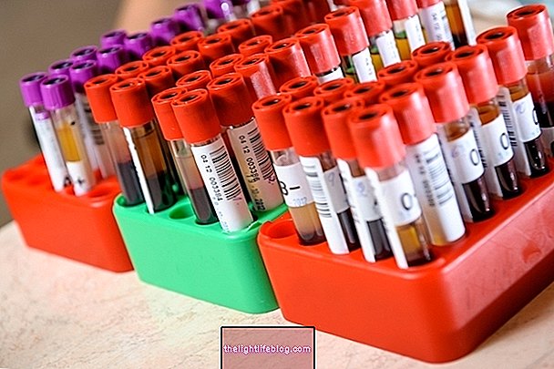
In addition, the obstetrician will also indicate blood tests to assess the levels of the hormone Beta-HCG, which in these cases are in very high amounts and if you suspect other diseases, you may recommend other tests such as urine, CT scan or MRI.
Treatment options
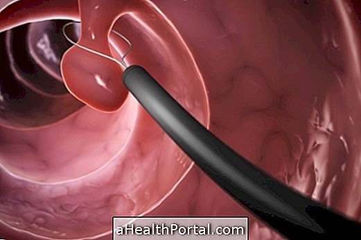
The treatment of molar pregnancy is based on performing a procedure called curettage, which consists of sucking the inside of the uterus to remove the abnormal tissue. In rarer cases, even after curettage, the abnormal cells can remain in the uterus and give rise to a type of cancer, called gestational choriocarcinoma and, in these situations, it may be necessary to have surgery, use chemotherapy drugs or undergo radiotherapy.
Furthermore, if the doctor finds that the woman's blood type is negative, she may indicate the application of a medicine, called matergam, so that specific antibodies do not develop, avoiding complications when the woman becomes pregnant again, such as fetal erythroblastosis, for example. Learn more about fetal erythroblastosis and how treatment is performed.
Was this information helpful?
Yes No
Your opinion is important! Write here how we can improve our text:
Any questions? Click here to be answered.
Email in which you want to receive a reply:
Check the confirmation email we sent you.
Your name:
Reason for visit:
--- Choose your reason --- DiseaseLive betterHelp another personGain knowledge
Are you a health professional?
NoMedicalPharmaceuticalsNurseNutritionistBiomedicalPhysiotherapistBeauticianOther
Bibliography
- UNIVERSITY OF PENNSYLVANIA. What is Gestational Trophoblastic Disease (GTD)?. Available in: . Accessed on 02 Dec 2019
- ANDRADE, Jurandyr M. Hydatidiform mole and gestational trophoblastic disease. Rev Bras Ginecol Obstet. Vol.31, n.2. 94-101, 2009
- CANDELIERA, Jean-Jacques. The hydatidiform mole. Cell Adh Migr. VOL.10, N.1-2. 226-235, 2016
- AMERICAN CANCER SOCIETY. Signs and Symptoms of Gestational Trophoblastic Disease. 2017. Available at:. Accessed on 03 Dec 2019


