Coats disease is a relatively rare disorder that affects the normal development of the blood vessels of the eye, more specifically the retina, where the images we see are created.
In people with this disease, it is very common for retinal blood vessels to rupture and, as a result, blood collects and causes inflammation of the retina, resulting in blurred vision, decreased vision, and in some cases even blindness.
Coats disease is more common in males and after 8 years of age, but can occur in anyone, even if there is no history of the disease in the family. Treatment should be started as soon as possible after diagnosis to avoid blindness.
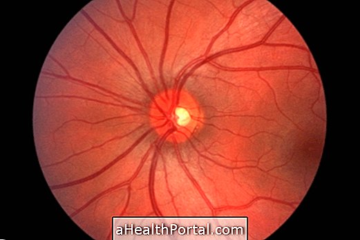
Main symptoms
Early signs and symptoms of Coats disease usually arise during childhood and include:
- Strabismus;
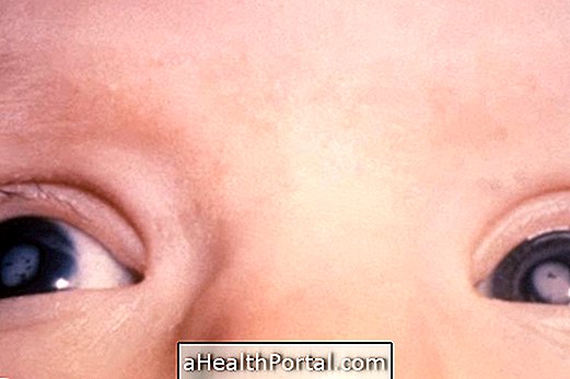
- Presence of a whitish film behind the lens of the eye;
- Decreased depth perception;
- Reduction of vision.
As the disease progresses, other symptoms may begin to appear, such as:
- Reddish color in the iris;
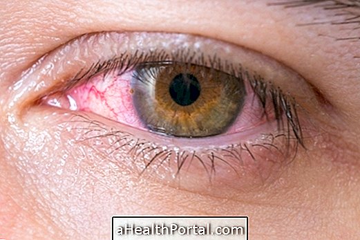
- Constant redness of the eye;
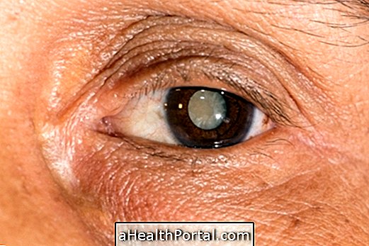
- Cataratas;
- Glaucoma.
In most cases, these symptoms affect only one eye, but they can also appear in both. Thus, whenever eye or vision changes occur, lasting more than a week, it is very important to see an ophthalmologist, even if they are affecting only one eye.
Who has a higher risk of having the disease
Coats disease can occur in anyone because it does not appear to be related to any genetic factor that can be inherited. However, it is more common in males and between the ages of 8 and 16, especially when there are symptoms of the disease until age 10.
How is the diagnosis made?
The diagnosis should always be made by an ophthalmologist through eye examination, assessment of eye structures and observation of symptoms. However, since the symptoms may be similar to those of other diseases of the eye, it may also be necessary to perform diagnostic tests such as retinal angiography, ultrasound or computed tomography, for example.
What are the stages of evolution?
The progression of Coats disease can be divided into 5 main stages:
- Stage 1 : There are abnormal blood vessels in the retina, but they are not yet ruptured and so there are no symptoms;
- Stage 2 : rupture of blood vessels of the retina occurs, leading to blood accumulation and gradual loss of vision;
- Stage 3 : retinal detachment occurs due to fluid accumulation, resulting in signs such as flashes of light, dark spots in the eye and discomfort in the eye. Learn more about retinal detachment;
- Stage 4 : With the gradual increase of fluid inside the eye, there is an increase in pressure that can result in glaucoma, in which the optic nerve is affected, greatly impairing vision. Here's how to identify glaucoma.
- Stage 5 : It is the most advanced stage of the disease when blindness and intense pain appear in the eye, due to the exaggerated increase of the pressure.
In some people, the disease may not progress through all phases and the time of evolution is quite variable. However, it is best to always start treatment when the first symptoms appear, to avoid the onset of blindness.
Treatment Options
Treatment is usually started to prevent worsening of the disease, so it should be started as soon as possible to prevent the onset of serious injuries leading to blindness. Some of the options that may be indicated by the ophthalmologist include:
1. Laser surgery
It is a type of treatment that uses a beam of light to shrink or destroy the abnormal blood vessels of the retina, preventing them from rupturing and leading to blood accumulation. Usually, this surgery is done in the earliest stages of the disease in the doctor's office and under local anesthesia.
2. Cryotherapy
In this treatment, instead of using a laser, the ophthalmologist makes small applications of extreme cold near the blood vessels of the eye to heal and close, preventing them from breaking.
3. Injection of corticosteroids
Corticosteroids are used directly in the eye to decrease inflammation in more advanced cases of the disease, helping relieve discomfort and may even improve vision. These injections need to be done in the doctor's office under local anesthesia.
In addition to these options, if there is retinal detachment or glaucoma, treatment should also be started for each of these consequences, in order to avoid aggravation of the lesions.

